Research
Winckler Lab
The field’s ignorance of the neuronal endosomal system constitutes a significant barrier to progress in several disease-oriented fields because the particular organization of the endosome determines postendocytic trafficking and thereby specific functional outcomes.
- Project 1: Discovery of neuronal adaptations to endosome organization and dynamics in dendrites.
-
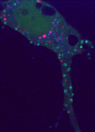
The field’s ignorance of the neuronal endosomal system constitutes a significant barrier to progress in several disease-oriented fields because the particular organization of the endosome determines postendocytic trafficking and thereby specific functional outcomes. Work with translational goals using disease-relevant cargos is still often carried out in non-neuronal cell types because the neuronal specializations in endosomal organization are underappreciated, and the insights gained from studying endosomes in non-neuronal cells do not translate into neurons. We thus set out to discover the organization and regulation of somatodendritic endosomes. In order to carry out this analysis, we mastered high-resolution live imaging of single endosomes, which allows for monitoring of dynamic behaviors, such as presumptive fusion and budding events, as well as loss and recruitment of cytosolic proteins to look at endosome maturation.
- Project 2: Mechanisms of sensing and responding to lysosomal stress in neurons.
-
There is a big knowledge gap for how neurons monitor and handle proteostatic stress, and how axons and dendrites might have adapted different mechanisms to do this effectively. One cause of proteostatic stress is lysosomal damage which occurs much more frequently than previously realized. Agents of lysosomal damage include lysosomotropic drugs (such as chloroquine and SSRIs), neurotoxic aggregates (such as a-synuclein, Ab, and tau), and oxidative stress (as occurs after ischemic stroke). Lysosomal damage results in lysosomal membrane permeabilization with immediate collapse of pH and calcium gradients, progressing to actual holes in the lysosomal membrane. Even though lysosomes are critical to neuronal function and often causally linked to neurological pathologies, the response of neurons to lysosomal damage on a mechanistic cellular level is poorly understood.
- Project 3: Signaling roles of intermediate filament isoforms in neural development.
-
While studying the trafficking of the cell adhesion molecule neurofascin, we discovered a new role for the Lissencephaly-linked doublecortin protein (DCX): DCX regulates the surface distribution of neurofascin by increasing its endocytosis. This is a novel role for DCX. Current models for the cellular actions of DCX place microtubules as the central effector for DCX. Importantly, the novel endocytic role of DCX does not require microtubule-binding of DCX. Our work thus opens a new avenue for studying and understanding additional roles for DCX in neurodevelopment and in lissencephaly. We are currently investigating non-microtubule roles for DCX using patient alleles of DCX, as well as analyzing new binding partners of DCX identified in a mass spectrometry screen. Our most recent work discovered the intermediate filament protein nestin as interacting with DCX. Surprisingly, nestin plays a signaling role downstream of Sema3a to modulate phosphorylation of DCX by cdk5.
- Project 4: Extracellular vesicles as neurotrophic signals in neurodevelopment
-
We are collaborating with Dr. Christopher Deppmann in the Biology Department at the University of Virginia who is an expert in nerve growth factor (NGF) signaling in the peripheral nervous system. Our interests and expertise in the cell biology of neuronal endosomes combined with Dr. Deppmann’s expertise in neurotrophin biology creates unique synergies. We discovered that NGF signaling endosomes come in multiple endosomal “flavors” and that a pathway discovered by the Deppmann lab, namely retrograde transcytosis, is responsible for generating this molecular diversification of signaling endosomes in dendrites. We discovered that sympathetic neurons in culture secrete extracellular vesicles (EVs). A subset of these EVs contains the NGF receptor TrkA. EV biology is a nascent field, but a range of EV functions have been described mainly in non-neuronal systems. EVs have a demonstrated role in tissue repair, immune surveillance, transportation of miRNAs, and activation of signaling cascades. Although there have been a handful of recent EV studies focusing on neurons, EVs have not previously been shown to participate in trophic neurodevelopmental processes.

